
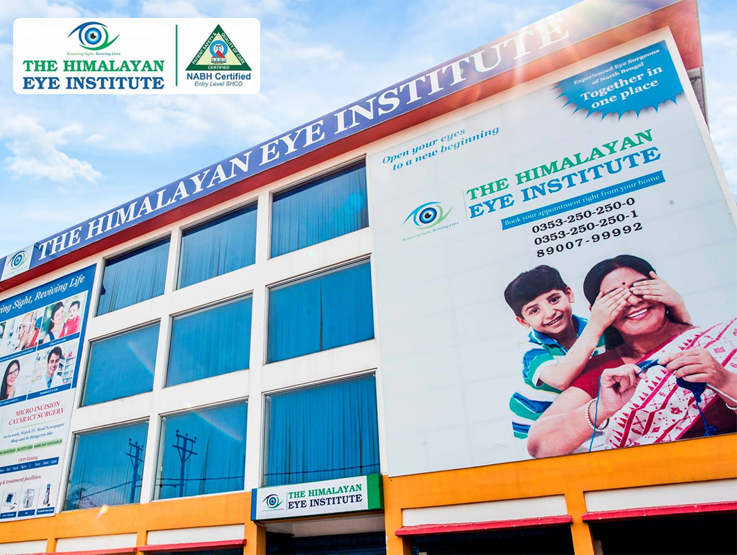
The Himalayan Eye Institute is the largest private eye service provider in North Bengal delivering high quality comprehensive eye services. It has received National Accreditation Board for Hospitals & Healthcare Providers (NABH) Entry Level certification in April 2022.
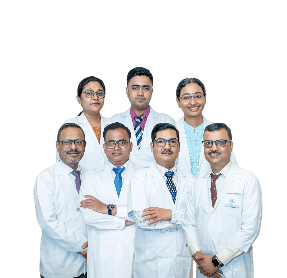
The hospital medical team is a group of skilled professionals who excel in their clinical and surgical skills. The team is made up of doctors who are highly respected in their fields for their expertise and know-how. Read More

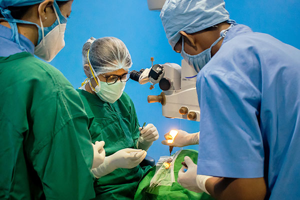
India has the world's largest blind population. About 90% of vision loss is preventable or treatable. Do not delay your eye check-up.

We bring variety of specs frames with current style and comfort. We also provide wide range of lenses including Blue light filter lenses, Scratch-Resistant coated Lenses, Anti-Reflective (ARC) Lenses, Photo chromatic or Day-Night Lenses that match with your lifestyle and requirement.
Our optical dispensing team uses cutting edge technology for accurate fitting of lenses to ensure high level of customer satisfaction. In addition, you will get Low Vision Aids, Ocular Prosthesis & Contact Lens services - all at one place!.
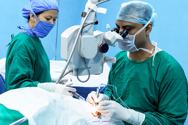
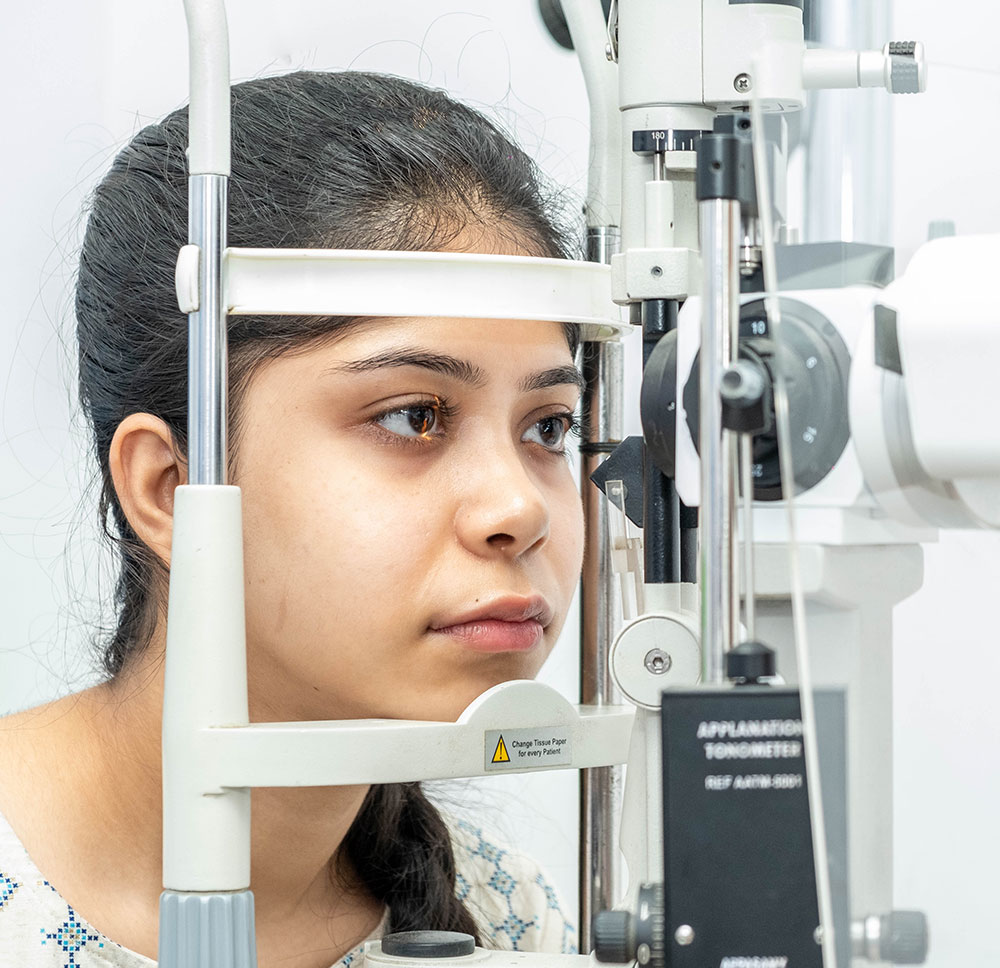
Read More...LASIK may not be appropriate for everyone! A Pentacam screening test can help you to determine whether you should go for LASIK or Phakic IOL. The Himalayan Eye Institute brings The OCULUS Pentacam®, the Gold Standard in anterior eye segment tomography. Besides Pentacam, the advanced ophthalmic diagnostics at the hospital includes Optical biometry, 3D OCT, HFA (perimetry), Specular Microscopy, FFA etc.


Looking for a second medical opinion to confirm a diagnosis or discuss alternative treatment options? Our International Patient's Services Desk is happy to provide you with travel documentation for visas, travel planning and also guide you to comfortable accommodation options near our hospitals. You may send us your reports and case history in advance, so that we can connect you with appropriate doctors and schedule an appointment.
 4.8 Rating |
 Today, my mother admitted in The Himalayan Eye Institute and completed Eye phaco . All the employees and doctors are very good attitude and coprative . Wishing the very glouries future. Suggest everyone for eye treatment. I think, the Himalayan Eye Institute is great for Eye Treatme

The quality eye care facility in Siliguri. Well maintained eye hospital. Taking utmost care post surgery patient by the doctors and by the nurses. Please visit if you have any eye related problems.

Very friendly staff , good hospitality and clean environment. Keep it up

Today my father's Surgery is done by Dr Shyamal Saha. Overall hospital facilities is great.

Cataract surgery done by Dr. Swarup kr. Roy. We, the family members of Panchali Basu(operated upon), are fully satisfied.

I have to visit the HEI frequently for my wife's eye treatment. Our experience every time has been very good. The standard maintained must be kept up, if not further improved upon. There's no end to betterment of a facility.

We visited the eye hospital and my father's cataract operation was done here. It's successful and all the services of the hospital are very satisfactory.

It's my first time in Himalayas eye institute All workers and Dr with whom am dealing with is awesome I really like the environment too Very happy with service Thanks to all

Best hospital. Good behavior among the staff & nurses. Went for cataract operation for the second eye. Excellent atmosphere.

I had accompanied my uncle for his checkup to the retina department today and the service provided by the receptionist and all the medical personnels was prompt. They were very helpful in all aspects. Moreover they guided us in each and every step. What I found wonderful is that they value money.. The Dr's charge as well as the investigations fees was very reasonable.

Copyrights © 2024, The Himalayan Eye Institute.