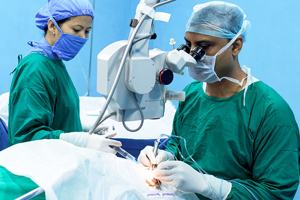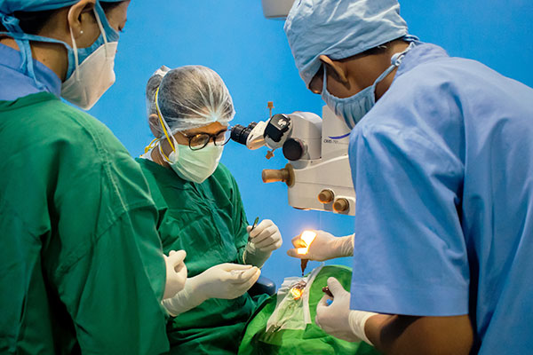

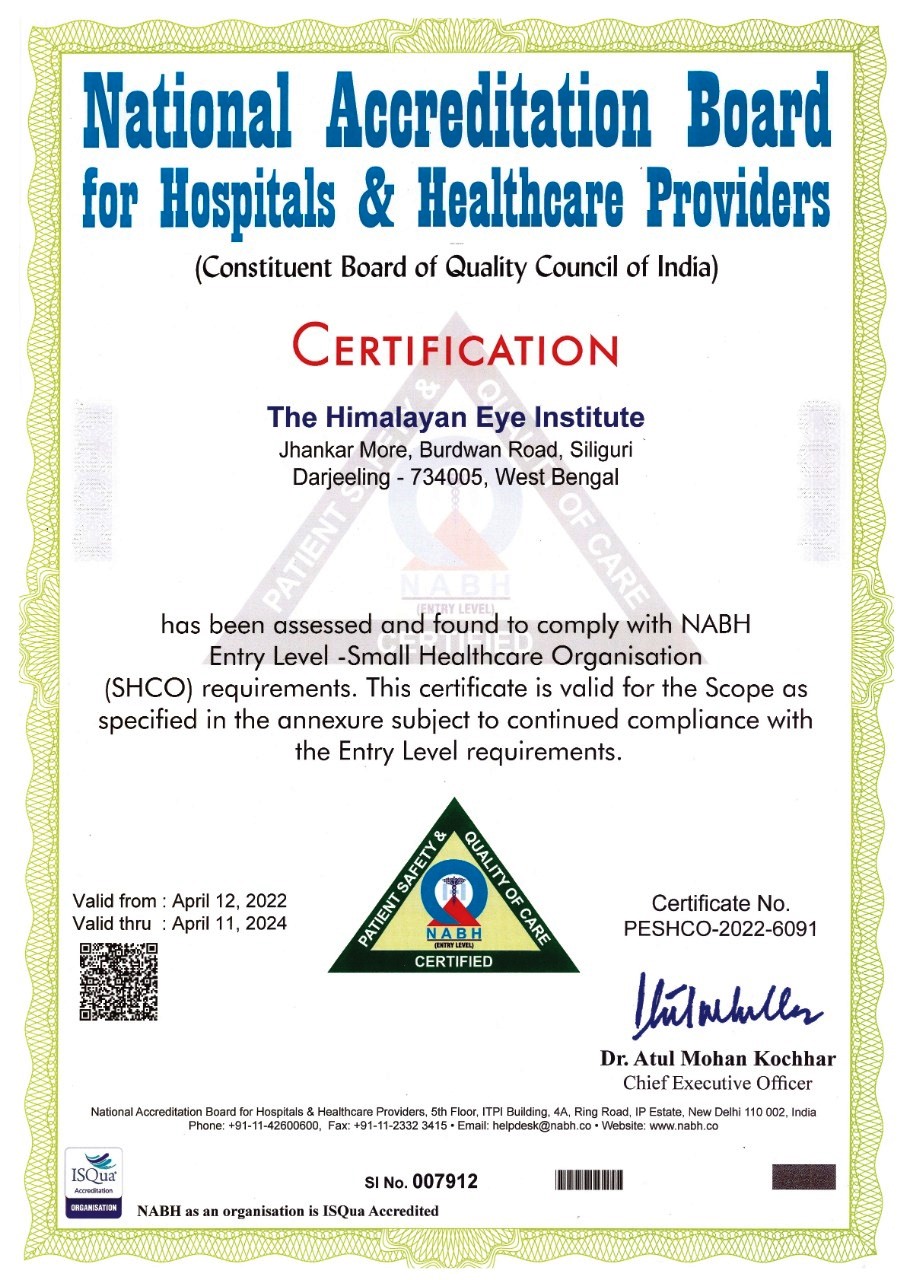
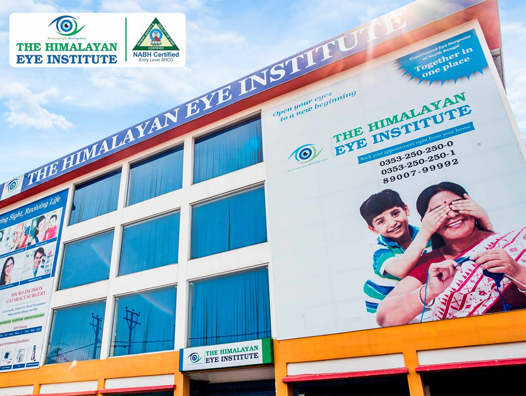
The Himalayan Eye Institute is the largest private eye service provider in North Bengal delivering high quality comprehensive eye services. It has received National Accreditation Board for Hospitals & Healthcare Providers (NABH) Entry Level certification in April 2022.
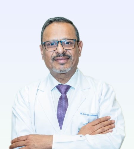
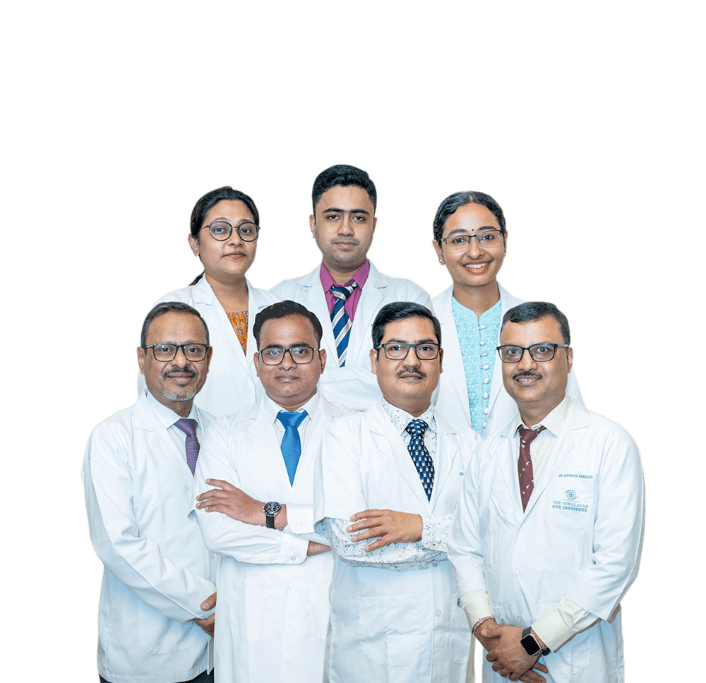
The hospital medical team is a group of skilled professionals who excel in their clinical and surgical skills. The team is made up of doctors who are highly respected in their fields for their expertise and know-how. Read More

India has the world's largest blind population. About 90% of vision loss is preventable or treatable. Do not delay your eye check-up.

We bring variety of specs frames with current style and comfort. We also provide wide range of lenses including Blue light filter lenses, Scratch-Resistant coated Lenses, Anti-Reflective (ARC) Lenses, Photo chromatic or Day-Night Lenses that match with your lifestyle and requirement.
Our optical dispensing team uses cutting edge technology for accurate fitting of lenses to ensure high level of customer satisfaction. In addition, you will get Low Vision Aids, Ocular Prosthesis & Contact Lens services - all at one place!.
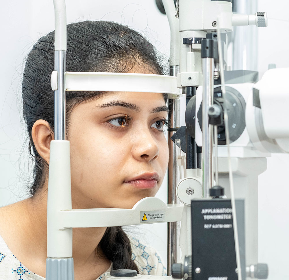
Read More...LASIK may not be appropriate for everyone! A Pentacam screening test can help you to determine whether you should go for LASIK or Phakic IOL. The Himalayan Eye Institute brings The OCULUS Pentacam®, the Gold Standard in anterior eye segment tomography. Besides Pentacam, the advanced ophthalmic diagnostics at the hospital includes Optical biometry, 3D OCT, HFA (perimetry), Specular Microscopy, FFA etc.


Looking for a second medical opinion to confirm a diagnosis or discuss alternative treatment options? Our International Patient's Services Desk is happy to provide you with travel documentation for visas, travel planning and also guide you to comfortable accommodation options near our hospitals. You may send us your reports and case history in advance, so that we can connect you with appropriate doctors and schedule an appointment.
 4.8 Rating |
 Very beautiful arrangement... I am very happy .. Very good doctor nurse n stuffs....

Excellent service is provided and behavior of the staffs is also appreciable. Place is very clean.

Very professional care and the overall management of the institute is well appreciated. Cleanliness and care is always reflected.

Very good helpful . Polite services. Management is very promising and prompt. Clean airy specious ⬇️ ⬆️ views all floors. Large screen photography showing from doctor's chambers. Happy with the treatment. Billing transparency quite impressive. No hidden charges.

Today we did my mother's operation from the Himalayan Eye Center hospital, Siliguri.. the overall experience is really good, all the doctors and supporting staffs are really helpful. Will definitely suggest everyone to visit here if facing any eye troubles.

Overall process including the Doctor and all employees are very cordial and friendly.

All the Doctors , Optometrists and other support stuffs are really very good, supportive and caring. The Institute maintains a good discipline of cleanliness, hygiene and all other Covid-19 norms. Medical Facilities available are almost at par with some Institutes outside West Bengal. Definitely the best place for eye check-up and treatment.

Recently my mom's eye surgery has done in this hospital the service of this hospital is very satisfying and cost is also reasonable i would recommend to all

Date: 6 Sept, 2022 Today we did my mother's operation from the Himalayan Eye Center hospital, Siliguri.. the overall experience is really good, all the doctors and supporting staffs are really helpful. Will definitely suggest everyone to visit here if facing any eye troubles.

All the facilities regarding treatment in the Retina dept have been excellent. The reception of staff is amiable..

Copyrights © 2024, The Himalayan Eye Institute.